Proctology – Pilonidal sinus

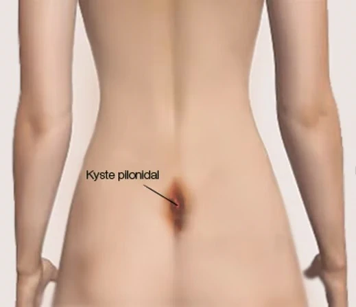
A pilonidal sinus, also called a pilonidal cyst or sacrococcygeal cyst, consists of hairs trapped under the skin in the subcutaneous fat, causing an inflammatory reaction or a secondary infection. Its size varies over time; it is tender, or even very painful in the case of an abscess, and it is always located in the same place, in the upper part of the cleft between the buttocks.

Several theories can explain how it develops:

The congenital theory: a defect in the migration of embryonic cells.

The acquired theory: friction on the skin in the sitting position is thought to cause the hairs to become embedded.

As this small subcutaneous cyst progresses, it leads to secondary infection (abscess) and/or fistulisation (the appearance of one or more openings that give rise to a discharge). Successive flare-ups of infection can then create new tracts, new abscess pockets and new midline openings in the cleft between the buttocks.

All pilonidal sinuses, whether or not they have been complicated by an abscess, are an indication for surgery, which consists of complete excision of the lesions. Depending on the case, the wound will be left open (and cared for by a home nurse while it heals) or closed (if the excision is small). The operation is performed under general anaesthesia. Day surgery or a 24-hour hospital stay is usual.
What is a sacrococcygeal cyst?

A “pilonidal sinus”, “sacrococcygeal cyst” or “pilonidal cyst” is a relatively common acute or chronic suppurative condition in young adults.

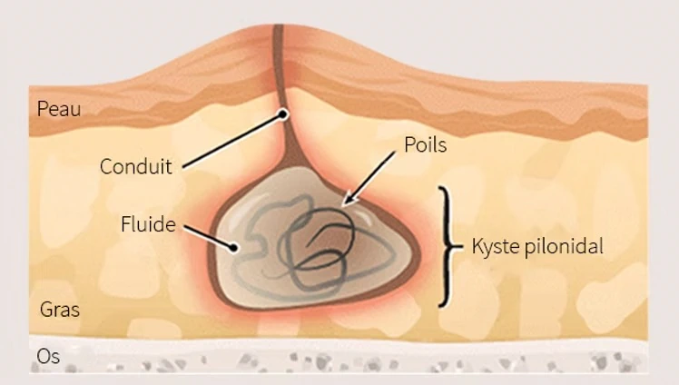
It is a pseudocystic cavity, sometimes containing hairs, located in the subcutaneous tissue, with a granulomatous wall; it is most often situated in the cleft between the buttocks and communicates with the skin through small channels that open via tiny holes on the midline: the pits.

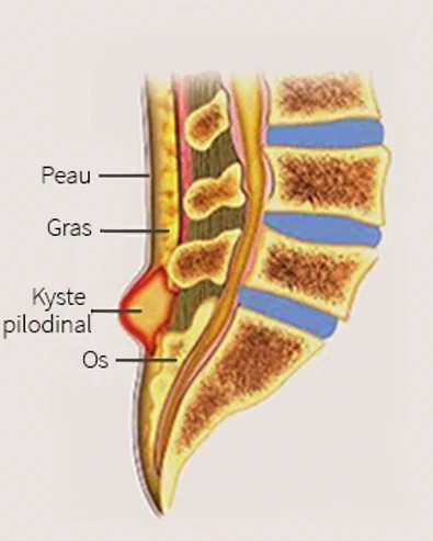
The pilonidal sinus is incorrectly called a “sacrococcygeal cyst” because it is usually located in the cleft between the buttocks (but it has no connection with the sacrum or the coccyx, nor does it have a lining of its own).

The exact frequency of pilonidal sinus is very difficult to assess; during the Second World War and the Korean War, it was so common among young American soldiers that it was called “Jeep disease”.
It should be noted that although the cleft between the buttocks is by far the most common location, it is not the only one: the sinus may be located at the front, on the anterior median raphe, in about 5% of cases.

Location of the pilonidal sinus in the sacrococcygeal region

Doctor, should I have surgery?

The answer is yes. Once a pilonidal sinus has formed, it never heals on its own.
Without treatment, it inevitably progresses, alternating between quiet phases and painful, sometimes severe, episodes.

How does the condition progress?
A pilonidal sinus typically follows a cycle of three phases:
Acute inflammatory flare-ups are the most distressing episodes: a red, hot and very painful hardened area appears in the cleft between the buttocks and may develop into an abscess requiring emergency drainage.
Quiescent phases are the calm periods: the patient has no symptoms, but the cavity is still present and relapse is a matter of time.
Chronic discharge phases involve seropurulent discharge in varying amounts, which is generally not very painful but troublesome in daily life.

What is the treatment?
Surgery is the only definitive treatment. The standard technique is complete “open” excision of the pilonidal cavity: the wound is not closed immediately, which allows gradual healing from the depths towards the surface and from the edges towards the centre. This results in a discreet linear scar in the cleft between the buttocks.
This healing is slow, generally two to three months, and requires regular dressing changes.
When the anatomy allows it, additional surgical techniques make it possible to close the wound partially, thereby appreciably reducing the duration and frequency of post-operative care.

Proctology, pilonidal sinus, Dr Deroide, Paris

How is the operation performed?

The operation can be performed under general anaesthesia or regional anaesthesia as same-day surgery, with discharge home the same evening. There is no special diet to follow.

Removing hair from the area of the cleft between the buttocks is advisable. It is important to empty the bladder before the operation to limit any difficulty passing urine after surgery. The operation takes about twenty minutes.

As with any surgical procedure, there are risks related to both the anaesthesia and the surgery, but they nevertheless remain fairly exceptional, particularly as regards post-operative bleeding.

The wound will be left open with packing inside it, which will be changed regularly by a nurse. The wound will then heal in a few weeks, and sometimes in a few months.

Sometimes, the wound will be fully closed with stitches that dissolve in about 10 days. With this option, care will be much simpler and quicker, provided the wound does not reopen, which can happen in about 50% of cases.

Post-operative advice

A return to normal life is possible straight away. Patients ideally go home on the evening of the operation, or otherwise the following morning. Painkillers are not always necessary; as a rule, it is not a very painful operation. The return to work depends on each person’s occupation, with an average of 10 days off work.

Physical activity will depend on how quickly the wound heals and can be resumed 4 weeks after the operation. Swimming in salt water or fresh water will only be possible once healing is complete, i.e. after 8 to 10 weeks on average.

Local care mainly consists of dressings. When the wound has been fully closed with stitches that dissolve in about 10 days, the dressings that need changing are very simple, and the wound can be washed in the shower.

Complete healing is expected around the end of the second week. In about 50% of cases, the wound may reopen (most often between the 3rd and 5th post-operative days).

The warning signs that the wound is reopening are increasing pain with discharge and redness. The stitches should then be cut to reopen the wound so that it can be packed as if it had been left open from the start.

The 2 advantages of this “closed” method are that in 50% of cases very little dressing care is needed (less than 15 days), and in the remaining 50%, as the wound has been closed in several layers, it usually reopens only in the superficial layers, which saves time compared with a wound left completely open. Dressings will be changed every 24, 48 or 72 hours depending on how much fluid the wound produces.

When the wound is left open and packed, this is known as healing by secondary intention. It is the simplest method for treating superficial tissue loss. It is a simple, inexpensive technique that relies solely on repeated dressings and requires a few basic rules to be followed.

This method requires regular follow-up of the patient, and therefore the patient’s cooperation. The results are, in principle, very satisfactory from a mechanical, functional and cosmetic point of view.

Healing goes through three phases. The first, known as the debridement phase, lasts eight to fifteen days. The aim of the debridement phase is to keep in check the natural infection present in the wound and to remove residual non-viable tissue. Bacteria take care of removing the remaining necrotic tissue.
The aim is to obtain a healthy, living base in the wound, known as granulation tissue. The duration of the second phase, known as the granulation phase, depends on the size of the cavity and on the absence of local secondary infection.
This phase can last several weeks. The granulation tissue will support the regrowth of skin from the wound edges, which corresponds to the third phase, known as epithelialisation: the concentric progression of the epidermal covering, which is the outermost layer of the skin.

Cross-sectional diagram of a pilonidal cyst under the skin

The principle of moist wound healing


Since the foundational studies of the 1960s, it has been established that healing is significantly faster in a moist environment than in a dry one, a discovery that revolutionised the design of modern dressings.
The figures speak for themselves: a wound kept moist under an occlusive dressing is more than 90% healed; left exposed to the air, it is only half healed; exposed to a current of warm air, the rate falls to just 18%.
However, moisture must remain balanced, neither insufficient nor excessive. This is precisely the role of modern advanced dressings, which regulate this environment using materials with specific properties.

Among the most widely used:
– Alginates, extracted from seaweed, absorb up to 10 to 15 times their weight in exudate. They turn into a gel on contact with the wound, trap debris, limit bacterial proliferation and have a haemostatic effect. Their removal is painless.
– Hydrofibres, which work in a similar way, share the same high absorbency.
– Hydrogels maintain the moisture needed to soften necrotic tissue, while alginates take over to absorb the excess when the wound is oozing.

How to change the dressings

The operation can be performed under general anaesthesia or regional anaesthesia as same-day surgery, with discharge home the same evening. There is no special diet to follow. Removing hair from the area of the cleft between the buttocks is advisable. It is important to empty the bladder before the operation to limit any difficulty passing urine after surgery. The operation takes about twenty minutes.
As with any surgical procedure, there are risks related to both the anaesthesia and the surgery, but they nevertheless remain fairly exceptional, particularly as regards post-operative bleeding.
The wound will be left open with packing inside it, which will be changed regularly by a nurse. The wound will then heal in a few weeks, and sometimes in a few months. Sometimes, the wound will be fully closed with stitches that dissolve in about 10 days. With this option, care will be much simpler and quicker, provided the wound does not reopen, which can happen in about 50% of cases.

PROBLEMS THAT MAY OCCUR

Frequently asked questions about pilonidal sinus

Book an appointment with Dr Deroide today for a personalised diagnosis.

Scroll to Top